Liver Biopsy Interpretation: Special Stains
What is a special stain?
Special stains are histochemical stains that have an affinity for particular extra/intra-cellular structures, or particular microorganisms.
What are the most common special stains used to assess liver biopsies?
All tissue slides are stained with hematoxylin and eosin (H&E). Hematoxylin stains nucleic acids (i.e. nuclei) a purple/blue color. Eosin stains components of the extracellular matrix/cytoplasm a pink color (Figure 1). Most liver biopsies are assessed using at least 2-3 H&E-stained slides, made from 4 um-thick tissue slices sectioned from sampled liver cores (~1.2 mm in thickness by a 16-gauge needle) at different depth .
Aside from standard H&E, the most common special stains used to assess liver biopsies include trichrome, iron, PAS-D, reticulin, and copper.
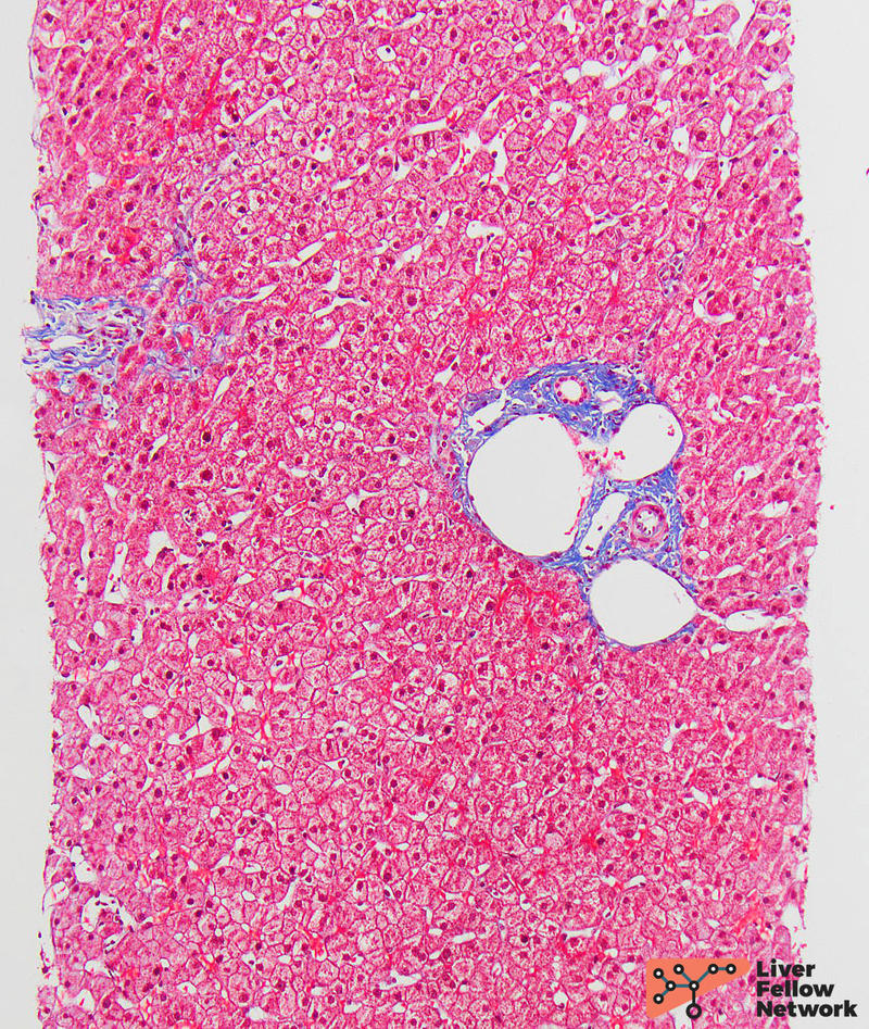
Trichrome is used to assess/stage fibrosis and it stains collagen blue. In normal liver biopsies there is normal collagen deposition around the portal tracts (Figure 2), while there is minimal deposition around the central veins. Patients with chronic liver disease often have increased fibrosis, which is assessed using one of the available scoring systems (IASL, Batts-Ludwig, and Metavir). The Batts-Ludwig is a commonly used system that stages fibrosis from 0-4: stage 0 (no fibrosis), stage 1 (fibrous portal expansion) (Figure 3), stage 2 (fibrosis extending beyond portal tracts with rare bridges or septae) (Figure 4), stage 3 (numerous bridges or septae) (Figure 5), and stage 4 (cirrhosis, regenerative nodules surrounded by fibrous bands) (Figure 6). Although stage 3 and early stage 4 fibrosis/evolving cirrhosis can appear similar, it is important to remember that in order to diagnose cirrhosis, the nodularity and bridging septa must involve the entire liver.
Perls iron is used to assess the level and distribution of iron deposition. In a normal liver biopsy, the iron stain is negative (Figure 7); however, in conditions with increased deposition, the iron appears as blue granules. These granules can be seen in patients with defects in iron metabolism (ex. hemochromatosis) (Figure 8) or in patients with secondary iron overload (ex. frequent blood transfusions) (Figure 9).
Periodic acid schiff (PAS) with diastase (PASD) is most often used in the assessment of alpha-1-antitrypsin (α1AT)-deficiency. A PAS stain highlights intrahepatic glycogen dark pink. In normal liver biopsies, when PAS is combined with diastase (PAS-D), the diastase digests out the glycogen and leaves the cytoplasm of hepatocytes a pale pink color (Figure 10). Additionally, in normal liver, PAS-D will stain lysosomes in Kupffer cells/macrophages. In contrast, patients with (α1AT)-deficiency harbor α1AT globules within hepatocytes that are resistant to the diastase, thus the globules remain dark pink even after digestion with the diastase (i.e. on PAS-D) (Figure 11).
Reticulin is used to assess liver architecture. It highlights the reticulin fibers (type III collagen) in the space of Disse, which helps to show the thickness of hepatocyte plates. Further, reticulin makes it easier to visualize areas of hepatocyte loss (collapse) or regeneration (increased thickness). In normal liver, the plates are 1-2 hepatocytes thick (Figure 12). Reticulin is used to distinguish well-differentiated neoplasms from one another, such as hepatocellular adenoma and hepatocellular carcinoma. It is also used in the diagnosis of nodular regenerative hyperplasia, as it shows the nodularity is caused by collapsed reticulin and not fibrosis (Figure 13).
Rhodanine is a special stain used to evaluate copper (copper associated protein) within hepatocytes. Normal hepatocytes show no increased copper deposition. In conditions with increased copper deposition, such as chronic cholestatic liver disease or Wilson's disease, the copper appears as orange-brown granules by rhodanine stain (Figure 14). It should be noted that in early stages of Wilson's disease, copper is diffusely distributed in the cytoplasm and often not detectable by histochemical stains. With disease progression, copper is sequestered within lysosomes and becomes visible by special stains in most (but not all) cases.
References
Clark I. and M. S. Torbenson, “Immunohistochemistry and special stains in medical liver pathology,” Advances in Anatomic Pathology. 2017, doi: 10.1097/PAP.0000000000000139.
- Saxena, Practical Hepatic Pathology: A Diagnostic Approach: Second Edition. 2017.


